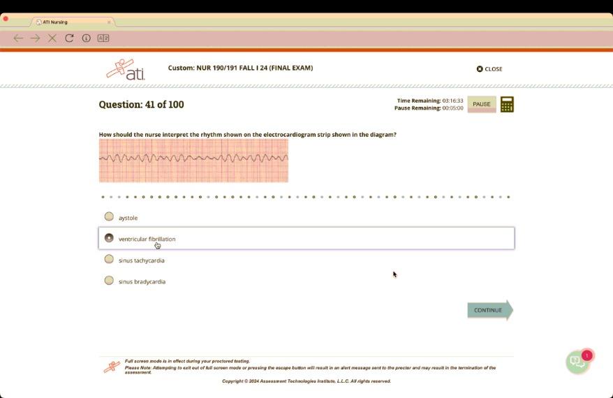
How should the nurse interpret the rhythm shown on the electrocardiogram strip shown in the diagram?
asystole
ventricular fibrillation
sinus tachycardia
sinus bradycardia
The Correct Answer is B
A. Asystole: Asystole would show a flat line with no electrical activity, which is not seen here.
B. Ventricular fibrillation: Ventricular fibrillation is characterized by chaotic, irregular waveforms without distinct P waves, QRS complexes, or T waves. The ECG strip shows this disorganized, erratic electrical activity consistent with ventricular fibrillation.
C. Sinus tachycardia: Sinus tachycardia would display a regular rhythm with identifiable P waves, QRS complexes, and T waves at a faster rate. This is not present in the ECG strip.
D. Sinus bradycardia: Sinus bradycardia would show a slower rate but with an organized rhythm and distinct P, QRS, and T waves. This is not indicated in the strip.
Free Nursing Test Bank
- Free Pharmacology Quiz 1
- Free Medical-Surgical Quiz 2
- Free Fundamentals Quiz 3
- Free Maternal-Newborn Quiz 4
- Free Anatomy and Physiology Quiz 5
- Free Obstetrics and Pediatrics Quiz 6
- Free Fluid and Electrolytes Quiz 7
- Free Community Health Quiz 8
- Free Promoting Health across the Lifespan Quiz 9
- Free Multidimensional Care Quiz 10
View Related questions
Correct Answer is B
Explanation
A. Early ventricular repolarization is represented by the T wave, not the P wave.
B. The P wave represents atrial depolarization, which is the electrical activity that triggers the contraction of the atria.
C. Slow repolarization of ventricular Purkinje fibers is represented by the T wave, not the P wave.
D. Ventricular depolarization is represented by the QRS complex, not the P wave.
Correct Answer is D
Explanation
A. Urinary frequency is characterized by the need to urinate more often but does not necessarily cause cloudy urine, odor, or hematuria.
B. Urinary retention involves the inability to empty the bladder fully but does not specifically present with cloudy urine, odor, or blood.
C. Urinary incontinence refers to the involuntary loss of urine and does not directly correlate with the urine's appearance or presence of blood.
D. A urinary tract infection (UTI) commonly causes cloudy urine, foul odor, and hematuria due to inflammation and infection in the urinary tract.