What is the rate on the rhythm strip in the image below?
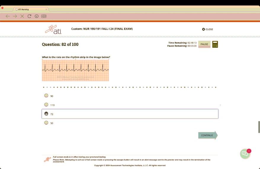
90
110
73
50
The Correct Answer is A
A. To determine the heart rate from a rhythm strip, you can count the number of R-R intervals in a set time frame (typically 6 seconds) and multiply by 10 to convert to beats per minute. If the rhythm is regular, you can also use the 300 method by dividing 300 by the number of large squares between R waves. In this case, the rate is calculated to be 90 beats per minute.
B. A heart rate of 110 beats per minute would be classified as tachycardia and is not supported by the observed intervals.
C. A heart rate of 73 beats per minute would be a normal resting heart rate but does not match the calculation from the rhythm strip.
D. A heart rate of 50 beats per minute would indicate bradycardia, which is not reflected in this rhythm strip.
Free Nursing Test Bank
- Free Pharmacology Quiz 1
- Free Medical-Surgical Quiz 2
- Free Fundamentals Quiz 3
- Free Maternal-Newborn Quiz 4
- Free Anatomy and Physiology Quiz 5
- Free Obstetrics and Pediatrics Quiz 6
- Free Fluid and Electrolytes Quiz 7
- Free Community Health Quiz 8
- Free Promoting Health across the Lifespan Quiz 9
- Free Multidimensional Care Quiz 10
View Related questions
Correct Answer is D
Explanation
A. A pustule is a small elevation of the skin that contains pus, typically smaller than 0.5 cm.
B. A macule is a flat, discolored area of skin that is less than 0.5 cm in diameter, so it does not fit the description of elevated lesions larger than 0.5 cm.
C. A papule is an elevated, solid lesion that is less than 0.5 cm in diameter; lesions larger than this would not be classified as papules.
D. A patch is defined as a flat, non-palpable lesion larger than 0.5 cm, and psoriasis can present as patches. Thus, the lesions described fit this classification.
Correct Answer is D
Explanation
A. Checking pupillary response to light assesses cranial nerve II (optic nerve).
B. Observing for facial symmetry primarily assesses cranial nerves VII (facial nerve) and possibly V (trigeminal nerve).
C. Testing for sense of smell assesses cranial nerve I (olfactory nerve).
D. Eliciting the gag reflex assesses cranial nerve IX (glossopharyngeal nerve) and also cranial nerve X (vagus nerve), making it the correct action to assess cranial nerve IX.