How should the nurse identify the rhythm shown in the diagram?
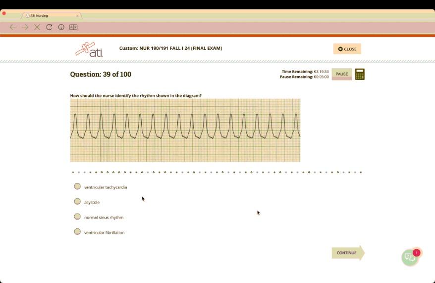
ventricular tachycardia
asystole
normal sinus rhythm
ventricular fibrillation
The Correct Answer is A
A. Ventricular tachycardia: Ventricular tachycardia (VT) is identified by a regular, fast rhythm with wide QRS complexes, typically without visible P waves. This rhythm often appears as consecutive, large, uniform waves, which is consistent with what is seen in the diagram.
B. Asystole: Asystole is characterized by a flat line, indicating no electrical activity, which is not present in this strip.
C. Normal sinus rhythm: Normal sinus rhythm would show identifiable P waves, a normal QRS complex, and a regular rate, which are not observed here.
D. Ventricular fibrillation: Ventricular fibrillation appears as chaotic, irregular waveforms with no clear QRS complexes or organization, which does not match the rhythm shown.
Free Nursing Test Bank
- Free Pharmacology Quiz 1
- Free Medical-Surgical Quiz 2
- Free Fundamentals Quiz 3
- Free Maternal-Newborn Quiz 4
- Free Anatomy and Physiology Quiz 5
- Free Obstetrics and Pediatrics Quiz 6
- Free Fluid and Electrolytes Quiz 7
- Free Community Health Quiz 8
- Free Promoting Health across the Lifespan Quiz 9
- Free Multidimensional Care Quiz 10
View Related questions
Correct Answer is C
Explanation
A. Metabolic alkalosis is characterized by a high pH and a high HCO3- level; this does not match the provided values.
B. Metabolic acidosis would show a low pH and a low HCO3-, which does not match the findings.
C. The pH is high (7.45) while the Paco2 is low (30 mm Hg), indicating respiratory alkalosis. The low HCO3- could be a compensatory mechanism but does not change the primary interpretation of respiratory alkalosis.
D. Respiratory acidosis would be indicated by a low pH and a high Paco2, which is not the case here.
Correct Answer is C
Explanation
A. The right upper quadrant is typically associated with gallbladder or liver issues, not duodenal ulcers.
B. The right lower quadrant is primarily associated with appendicitis or other conditions involving the appendix.
C. The left upper quadrant is where the duodenum is located, making it the appropriate area to assess for pain related to a duodenal ulcer.
D. The left lower quadrant is often associated with conditions affecting the sigmoid colon or left ovary but not typically with duodenal ulcers.