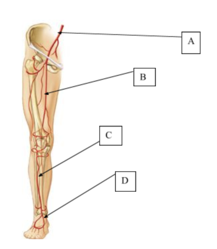
A nurse is assessing a client's peripheral circulation. In which of the following locations should the nurse palpate to assess the posterior tibial pulse? (Selectable areas, or "Hot Spots," are outlined in the artwork below. Select only the outlined area that corresponds to your answer.)
Iliac
Femoral
Popliteal
Tibial
The Correct Answer is D
A. The iliac pulse is located near the pelvis and is not used for assessing circulation in the lower extremities.
B. The femoral pulse is located in the upper thigh, not near the posterior tibial area.
C. The popliteal pulse is found at the back of the knee and is higher than the posterior tibial location.
D. The posterior tibial pulse is correctly located behind the medial malleolus on the inner side of the ankle. This location is where the posterior tibial artery is accessible and is commonly used to assess blood flow to the lower extremities.
Free Nursing Test Bank
- Free Pharmacology Quiz 1
- Free Medical-Surgical Quiz 2
- Free Fundamentals Quiz 3
- Free Maternal-Newborn Quiz 4
- Free Anatomy and Physiology Quiz 5
- Free Obstetrics and Pediatrics Quiz 6
- Free Fluid and Electrolytes Quiz 7
- Free Community Health Quiz 8
- Free Promoting Health across the Lifespan Quiz 9
- Free Multidimensional Care Quiz 10
View Related questions
Correct Answer is D
Explanation
A. The iliac pulse is located near the pelvis and is not used for assessing circulation in the lower extremities.
B. The femoral pulse is located in the upper thigh, not near the posterior tibial area.
C. The popliteal pulse is found at the back of the knee and is higher than the posterior tibial location.
D. The posterior tibial pulse is correctly located behind the medial malleolus on the inner side of the ankle. This location is where the posterior tibial artery is accessible and is commonly used to assess blood flow to the lower extremities.
Correct Answer is A
Explanation
A. +4 pitting edema is characterized by severe pitting that creates a deep indentation (greater than 8 mm) that remains for a prolonged period. This description matches the findings in option
B. This describes +1 pitting edema, which is not consistent with +4 edema.
C. This option describes +2 or +3 pitting edema, as the indentation subsides rapidly, which does not align with +4.
D. Although this describes deep pitting, the depth is less than 8 mm, which is not consistent with +4 edema.